
A comprehensive eye exam does two jobs. It finds the prescription that gives you the clearest, most comfortable vision, and it checks the health of your eyes for problems you may not notice yet. Many serious eye conditions, including glaucoma, cause no symptoms in their early stages.
Here is what a typical exam at Eye Q involves. Not every patient needs every test; the doctor tailors the exam to your age, history and symptoms.
Your history and concerns
The exam starts with a conversation: changes you have noticed, how you use your eyes at work and at home, medications, general health conditions such as diabetes or high blood pressure, and family history of eye disease. Mention headaches, eye strain, dryness or trouble with night driving, even if they seem minor.
Measuring your prescription
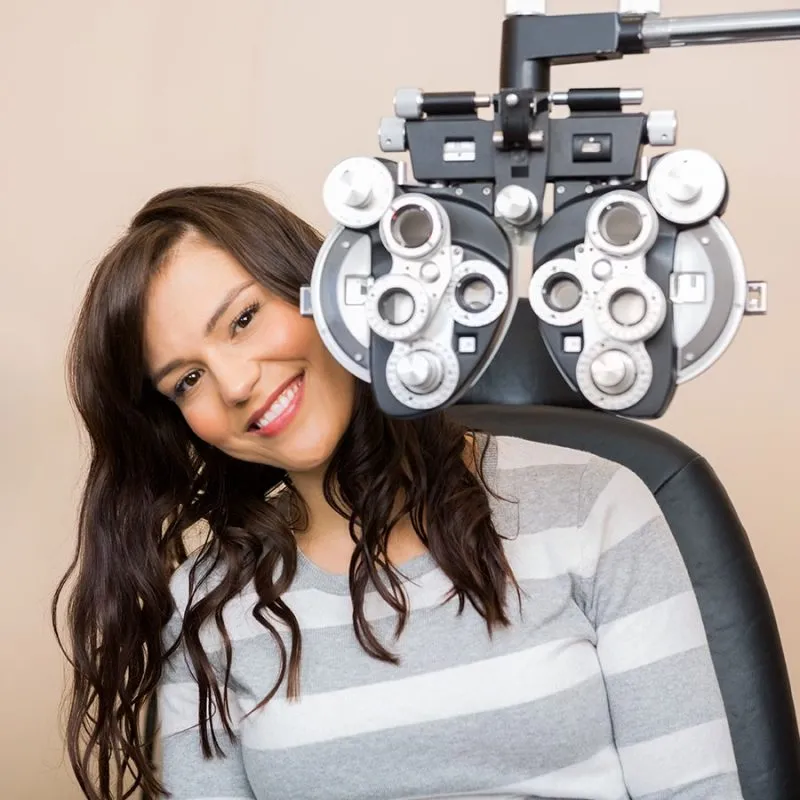
An automated measurement gives the doctor a starting point. The doctor then refines your prescription lens by lens, asking which option looks clearer. This is also when the doctor considers your daily needs, such as computer work, reading or sports, which can affect which lenses are recommended.
How your eyes work together
Clear vision in each eye is only part of the picture. The doctor checks eye alignment, focusing and how well the eyes team up, especially at reading distance. Problems here can cause fatigue, headaches or difficulty concentrating on close work.
Eye pressure and the front of the eye
A pressure measurement is one of the screening tests for glaucoma, a condition that damages the optic nerve gradually and painlessly. The National Eye Institute notes that early detection is the best way to protect vision. Using a microscope with a bright light, the doctor also examines the eyelids, cornea and lens for signs of dry eye, infection or cataracts.
Retinal imaging
Retinal photography captures a detailed image of the back of the eye: the retina, optic nerve and blood vessels. Comparing images from year to year makes subtle changes easier to spot. Retinal photos are especially useful for monitoring diabetic eye disease and macular degeneration.
The iWellness OCT scan
Eye Q uses iWellness, a type of optical coherence tomography (OCT). OCT uses light to create cross-sectional images of the retina and optic nerve, showing their individual layers. The American Optometric Association describes OCT as a noninvasive way to detect and monitor conditions such as glaucoma and macular disease.
The scan takes only moments, involves no contact with the eye and in most cases doesn't require dilation. It doesn't replace a dilated exam when one is needed, but it adds information that a standard view of the retina can't show.
Visual field testing
A visual field test maps your side vision by asking you to respond when lights appear in different places. Gaps in the field can point to glaucoma, optic nerve problems or certain neurological conditions. It is often used for people with risk factors or symptoms, and to monitor known conditions over time.
Dilation
Dilating drops widen the pupils so the doctor can examine more of the retina. Your vision may be blurry and sensitive to light for several hours afterward, so bring sunglasses and consider arranging a ride. Ask when you book whether dilation is likely at your visit.
Wrapping up
Before you leave, the doctor should explain what they found, whether anything needs monitoring or treatment, and when to come back. If you have a new prescription, our opticians can help you choose lenses and frames suited to how you use your eyes.
See what's included in comprehensive eye exams at Eye Q.